")
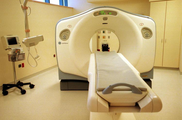
Radiation exposure from medical imaging equipment could be reduced by a factor of ten or more, according to a new report. Researchers at the University of California, San Diego (UCSD) say they have discovered an approach for processing X-ray data, utilizing video game processors.
This development follows years of concern from a number of medical groups, who warn of risks associated with staggering increases of radiation exposure to patients. For each year’s use of today’s scanning technology, the resulting cancers could cause about 14,500 deaths, according to authors of the UCSD report.
Meanwhile, use of radioactive imaging equipment continues to rise. A 2009 study from the U.S. National Council on Radiation Protection and Measurements found that Americans were exposed to seven times more radiation from medical procedures in 2006 than in 1980. In 2007, The New England Journal of Medicine estimated that the U.S. conducted at least 62 million CT (Computed Tomography) scans annually.
One of the reasons for frequent use of CT scans is that they can be read more quickly than other tests that do not use radiation, such as MRI’s, making it a convenient method in hospital emergency rooms when looking for the cause of abdominal or chest pains and headaches. Another reason for the increase stems from cancer treatments that employ repeated scans.
Cone beam CT, for example, plays an essential role in image-guided radiation therapy (IGRT), a state-of-the-art cancer treatment that utilizes repeated scans during a course of radiation therapy in an effort to precisely target tumors. However, the large cumulative radiation dose from the repeated scans has raised concerns among physicians and patients.
Earlier this year, the U.S. Food and Drug Administration (FDA) called for a reduction in radiation exposure from CT scans, as well as nuclear medicine studies, and fluoroscopy. According to the FDA, one CT abdomen scan is equal to about 400 chest X-rays.
“CT dose has become a major concern of the medical community. Our work, when extended from cancer radiotherapy to general diagnostic imaging, may provide a unique solution to solve this problem by reducing the CT dose per scan by a factor of 10 or more,” says Steve Jiang, senior author of the study and a UCSD associate professor of radiation oncology.
While it had previously been possible to reduce the total number of X-ray projections by tuning down the X-ray generator pulse rate during a CT scan to minimize a patient’s exposure to radiation, the option came at a price—mathematically incomplete data that takes hours to process.
However, with recent advances in the field of compressed sensing, the research team at UCSD was able to develop an innovative CT reconstruction algorithm for graphic processing unit (GPU) platforms. The GPU processes data in parallel, increasing computational efficiency and making it possible to reconstruct a cone beam CT scan in about two minutes.
This innovation represents a totally new application for GPU cards—devices that were originally designed to power 3-D computer graphics—especially for video games.
With only 20 to 40 in total numbers of X-ray projections and 0.1 mAs per projection, the research team achieved images clear enough for image-guided radiation therapy. The reconstruction time ranged from 77 seconds to 130 seconds on an NVIDIA Tesla C1060 GPU card, depending on the number of projections—an estimated 100 times faster than similar iterative reconstruction approaches.
Compared to the currently available and widely used scanning protocol of about 360 projections with 0.4 mAs per projection, the new processing method resulted in 36 times to 72 times less radiation exposure for patients, explains the report’s lead author and UCSD postdoctoral fellow, Xun Jia in a statement
“With our technique, we can reconstruct cone beam CT images with only a few projections—40 in most cases—and lower mAs levels,” Jia added. “This considerably lowered the radiation dose.”