Imagine you have shrunk to a microscopic size and you are standing on a molecule inside a human body.
The molecule is as big as a planet under your feet. The surrounding molecules are so far away from you that they are like the other planets that surround Earth—visible only through a telescope or as a dot in the night sky.
Viewed with the naked eye, the human body and all other objects appear solid. But take a minute to consider that your body is made of particles that are constantly moving, with spaces between them that could appear vast from a microscopic perspective.
No one has taken a photo from the surface of a molecule as described, so we'll leave that view up to your imagination. But here’s a close look at the human body through the lens of a microscope.
Blood Cells

(Bruce Wetzel, Harry Schaefer/National Cancer Institute)
A scanning electron microscope image shows circulating human blood. The image captures several white blood cells, red blood cells, lymphocytes, a moncyte, a neutrophil, and many disc-shaped platelets. The lymphocytes fight disease by producing antibodies. Platelets form in bone marrow and are necessary for blood clotting.
Nerve End

(Tina Carvalho/National Institutes of Health)
Retina (Part of the Eye)

(National Center for Microscopy and Imaging Research)
Skin
Cross section of a human skin cell.
Appendix
Cross section of a normal human appendix as seen with a light microscope at low magnification.
Cancer

(Tom Deernick via National Institutes of Health)
Hair Follicle
Spinal Cord Neuron
A silver stained spinal cord neuron.
Tongue

(National Center for Microscopy and Imaging Research)
Here are a couple bonus photos, not of the human body, but interesting microscopic views nonetheless.

(Dartmouth College/Wikimedia Commons)
A fruit fly’s compound eye.
Plant cells in which green chlorophyll can be seen.
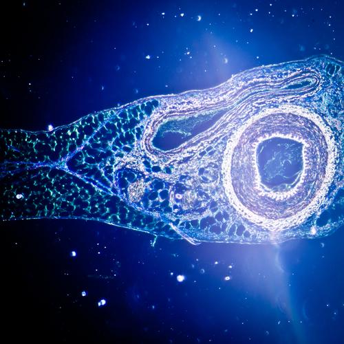
*Image of blood vessel via Shutterstock.


