CUHK Develops New Technique to Deeply Visualize 3D Structure of Brain’s Biological Tissue

CUHK Develops New Technique to Deeply Visualize 3D Structure of Brain’s Biological Tissue
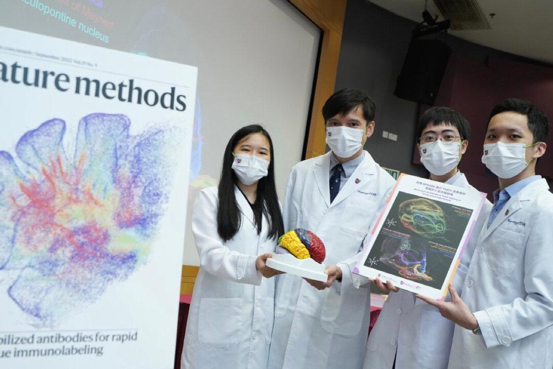
A team at the Margaret K.L. Cheung Research Centre for Management of Parkinsonism, Faculty of Medicine, CUHK, has developed a rapid and widely applicable biological staining technique to visualize the 3D structures of biological tissue, including the structure of the brain. (Courtesy of the Chinese University of Hong Kong)
9/26/2022
Updated:
3/16/2023

The technique of visualizing the 3D structure of the brain’s biological tissues is particularly important for understanding the pathological process of neurological diseases, such as, Parkinson’s and Alzheimer’s. However, the current technology which can fully visualize the brain structure is not widely used and is costly.

A team at the Margaret K.L. Cheung Research Centre for Management of Parkinsonism of the Chinese University of Hong Kong (CUHK), has developed a 3D immunostaining technique—ThICK (thermal immunohistochemistry with optimised kinetics) staining technology, which can produce stabilized antibodies for rapid labeling and imaging of molecules in biological tissue, and deeply visualize their 3D structure. The research findings were published as the cover story in the latest issue of Nature Methods, an international scientific journal.
Immunostaining, widely used to visualize the structures of biological tissues, uses antibodies to target biological molecules in tissue and show the structure of the tissue.

Dr. Lai Hei-ming, Assistant Professor in the Department of Psychiatry, Faculty of Medicine of CUHK, and Principal Investigator of the Margaret K.L. Cheung Research Centre for Management of Parkinsonism, pointed out that usually, antibodies are sensitive to heat and chemicals called “denaturants.”

When used in immunostaining, these properties limit the choice of temperature and denaturants, since they can destabilize the antibodies and make them lose their functions. The antibodies cannot reach deep into the tissues, hindering their application in high-throughput, 3D, structural studies.

The team developed a chemical technology that can convert antibodies, which are commonly used in research, making them much more resistant to heat and denaturants. The heat-stable antibodies termed SPEARs (synergistically protected polyepoxide-crosslinked Fab-complexed antibody reagents), can be used to achieve immunolabeling of biological molecules deep inside intact tissues.

Therefore, the use of SPEARs in ThICK staining can help to overcome the limitations of deep 3D immunostaining and optimize the visualization of brain structures.

Dr. Zachary Lau, the investigator of the Margaret K.L. Cheung Research Centre for Management of Parkinsonism, said that compared with other advanced technologies, ThICK staining achieves whole-organ immunolabeling faster, from requiring one to eight weeks for each round of antibody staining to a much shorter time of one to three days.

The research team used SPEARs to conduct the ThICK staining technology, and the results showed that it can achieve whole mouse brain immunolabeling within 72 hours. When applied to human brain tissues, it can achieve nearly fourfold deeper penetration with threefold fewer antibodies.

The research team said that this new method is compatible with a wide range of available antibodies, and different classes of tissue preservation and clearing methods. It can therefore be readily handled and implemented in most laboratories. This new technique is especially applicable to the study of neurodegenerative diseases.

The team hopes that their work can accelerate the path toward new tools for disease diagnosis and treatment.