Autoimmune uveitis is an ocular disease caused by inflammation of the tissues inside the eyeball, which can lead to permanent blindness. A research team from the Faculty of Medicine at the Chinese University of Hong Kong (CUHK) discovered that GHRH (Growth Hormone-Releasing Hormone), a biological pathway that induces immune choroiditis, can be inhibited using currently available drugs and is expected to become a new treatment means for the disease.
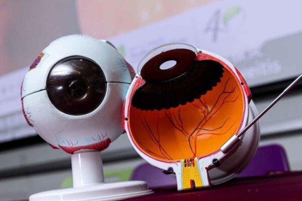
The team pointed out that the uvea is the inner tissue of the eyeball, located between the sclera and the retina. Because of its dark purple color, it is sometimes called the choroid. Infectious uveitis is caused by bacterial or viral infection. The patient’s lymphocytes often show abnormal responses to retinal antigens, which can evolve into an autoimmune disease in the long term.
Professor Clement Tham Chi-yung, Chairperson of the Department of Ophthalmology and Visual Sciences (DOVS), Faculty of Medicine at CUHK, explained that common symptoms of uveitis include red eyes, pain, and blurred vision. It is estimated that 150 out of 100,000 people in China suffer from uveitis every year. Autoimmune uveitis is a chronic, relapsing disease, and there is currently no effective cure.
Assistant Professor Simon Szeto Ka-ho of DOVS, added that steroids are a standard treatment for autoimmune uveitis, but they are not effective for some patients, and long-term use of high-dose steroids can lead to other complications, such as cataracts, glaucoma, among others. Second-line treatments have limited efficacy and also have many side effects. Biological agents are relatively expensive and may even lead to drug resistance.
The team used an experimental mice model to simulate patients with autoimmune uveitis and discovered a biological pathway that releases neuroendocrine growth hormone, the GHRH pathway. The signal received through transmission of the GHRH pathway induces the Th17 differentiation of pathogenic cells causing an imbalance, thus stimulating the production of inflammatory cytokines and evolving into autoimmune uveitis. The team also found that some registered drugs can inhibit the GHRH pathway and are expected to become new treatment means.
Dr. Chu Wai-kit, Research Assistant Professor of DOVS said that the research results show that while ocular and neuroinflammation are caused by Th17 cells, the GHRH receptor is an important regulator of Th17 differentiation of pathogenic cells. In other words, employing GHRH inhibitors can reduce the number of Th17 cells and thereby inhibit autoimmune uveitis. However, GHRH receptor inhibitors also exhibit the possibility of inhibiting normal growth and development, making clinical application difficult. However, the team has identified one downstream signaling pathway called JAK-STAT3 and found certain readily available drugs that can inhibit this pathway.


Du Lin, a postdoctoral researcher at DOVS, added that Th17 cells may cause other autoimmune diseases, including rheumatoid arthritis and multiple sclerosis, and based on the same theory, GHRH receptor inhibitors have the potential to suppress autoimmune neuroinflammation.